Fitxer:Wound healing phases.png
Fitxer original (6.338 × 1.236 píxels, mida del fitxer: 859 Ko, tipus MIME: image/png)
| Aquest fitxer i la informació mostrada a continuació provenen del dipòsit multimèdia lliure Wikimedia Commons. |
Resum
| Descripció |
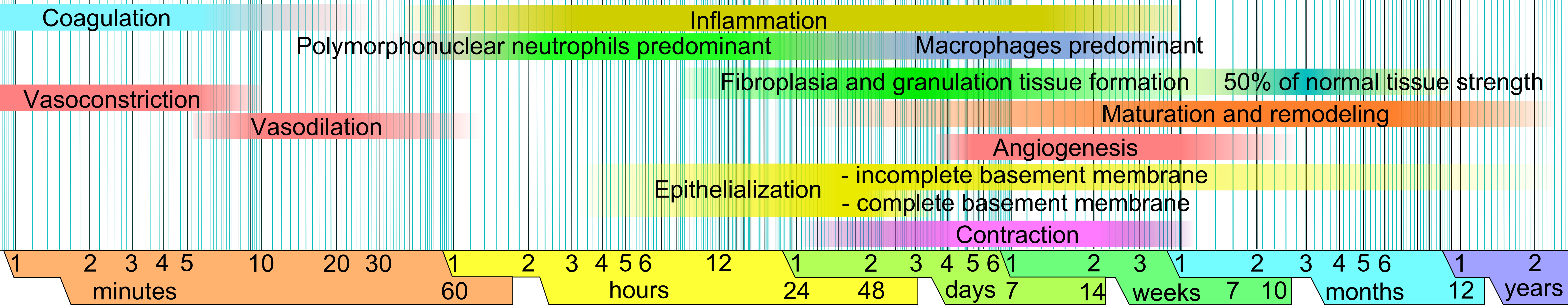
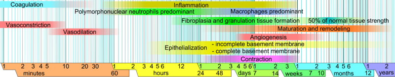
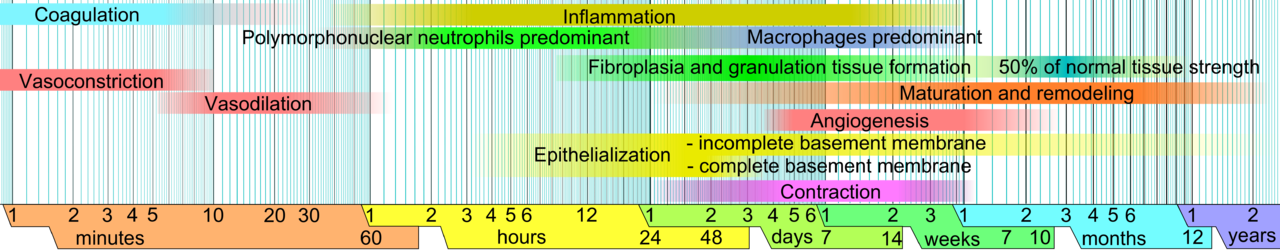
English: Phases of wound healing. Limits vary within faded intervals, mainly by wound size and healing conditions, but image does not include major impairments that cause chronic wounds. |
| Data | |
| Font | Treball propi (from the template Logarithmic time scale - milliseconds to years.svg) |
| Autor |
When using this image in external works, it may be cited as:
or
|
| Altres versions | العربيَّة |
References
The direct URL link to this reference list is: http://commons.wikimedia.org/wiki/File:Wound_healing_phases.png#References
Inflammation and upper limit of beginning of maturation and remodeling, as well as its ending:
- worldwidewounds.com > Figure 3 - The time relationship between the different processes of wound healing. by Gregory S Schultz, Glenn Ladwig and Annette Wysocki - in turn adapted from Asmussen PD, Sollner B. Mechanism of wound healing. In: Wound Care. Tutorial Medical Series. Stuttgart: Hippokrates Verlag, 1993.
Lower limit of beginning of maturation and remodeling, and equivalent limit for fibroplasia and granulation tissue formation:
- Fig. 9-1. The cellular, biochemical, and mechanical phases of wound healing. Pollock, Raphael E.; F. Charles Brunicardi; Dana Lynne Andersen; Billiar, Timothy R.; Dunn, David; Hunter, John G.; Matthews, Jeffrey J. (2009) Schwartz's Principles of Surgery, Ninth Edition, McGraw-Hill Professional ISBN: 0-07-154769-X.
Vasoconstriction and vasodilation:
- Stadelmann W.K., Digenis A.G. and Tobin G.R. (1998). Physiology and healing dynamics of chronic cutaneous wounds. The American Journal of Surgery 176 (2): 26S-38S. PMID 9777970 Hamilton, Ont. B.C. Decker, Inc. Electronic book
Angiogenesis:
- Nguyen, D.T., Orgill D.P., Murphy G.F. (2009). Chapter 4: The Pathophysiologic Basis for Wound Healing and Cutaneous Regeneration. Biomaterials For Treating Skin Loss. CRC Press (US) & Woodhead Publishing (UK/Europe), Boca Raton/Cambridge, p. 25-57. (ISBN 978-1-4200-9989-9 Invalid ISBN, ISBN 978-1-84569-363-3)
Polymorphonuclear neutrophils and ending of fibroplasia and granulation tissue formation:
- de la Torre J., Sholar A. (2006). Wound healing: Chronic wounds. Emedicine.com. Accessed January 20, 2008. http://www.emedicine.com/plastic/topic477.htm
Macrophages:
- Expert Reviews in Molecular Medicine. (2003). The phases of cutaneous wound healing. 5: 1. Cambridge University Press. Accessed January 20, 2008. http://www-ermm.cbcu.cam.ac.uk/03005829a.pdf
Upper limit of beginning of fibroplasia and granulation tissue formation (collagen deposition), epithelialization and contraction:
- Romo T. and Pearson J.M. 2005. Wound Healing, Skin. Emedicine.com. Accessed December 27, 2006.
Additional note on contraction:
- Mulvaney M. and Harrington A. 1994. Chapter 7: Cutaneous trauma and its treatment. In, Textbook of Military Medicine: Military Dermatology. Office of the Surgeon General, Department of the Army. Virtual Naval Hospital Project. Accessed through web archive on September 15, 2007. https://web.archive.org/web/20031218072356/http://www.vnh.org/MilitaryDerm/Ch7.pdf
Percentage of normal tissue strength:
- Mercandetti M., Cohen A.J. (2005). Wound Healing: Healing and Repair. Emedicine.com. Accessed January 20, 2008. http://www.emedicine.com/plastic/topic411.htm

|
File:Wound healing phases.svg és una versió vectorial (SVG) d'aquest fitxer. En cas de ser millor, hauria de ser emprada en lloc d'aquesta imatge tramada.
File:Wound healing phases.png → File:Wound healing phases.svg
Per a més informació pel que fa als gràfics vectorials, llegiu la transició a SVG en Commons. També hi ha informació quant a la compatibilitat del MediaWiki amb les imatges SVG. |
Llicència
| Jo, el titular del copyright d'aquesta obra, l'allibero al domini públic. Això s'aplica a tot el món. En alguns països això pot no ser legalment possible, en tal cas: Jo faig concessió a tothom del dret d'usar aquesta obra per a qualsevol propòsit, sense cap condició llevat d'aquelles requerides per la llei. |
Historial del fitxer
Cliqueu una data/hora per veure el fitxer tal com era aleshores.
| Data/hora | Miniatura | Dimensions | Usuari/a | Comentari | |
|---|---|---|---|---|---|
| actual | 07:24, 18 gen 2011 | 6.338 × 1.236 (859 Ko) | Mikael Häggström | Moved info in infobox to image page instead. | |
| 06:52, 14 nov 2010 | 6.338 × 1.236 (926 Ko) | Mikael Häggström | simplified legend | ||
| 06:49, 14 nov 2010 | 6.338 × 1.236 (933 Ko) | Mikael Häggström | another update | ||
| 17:05, 13 nov 2010 | 6.338 × 1.236 (900 Ko) | Mikael Häggström | Removed details about constituents. See sections on granulation tissue formation and remodeling for such details. | ||
| 16:07, 13 nov 2010 | 6.338 × 1.307 (957 Ko) | Mikael Häggström | changed succession order | ||
| 16:07, 13 nov 2010 | 6.338 × 1.307 (957 Ko) | Mikael Häggström | changed succession order | ||
| 15:53, 13 nov 2010 | 6.338 × 1.307 (949 Ko) | Mikael Häggström | minor adjustment | ||
| 15:47, 13 nov 2010 | 6.338 × 1.307 (946 Ko) | Mikael Häggström | Distinguished collagen types | ||
| 21:00, 3 nov 2010 | 6.444 × 1.209 (940 Ko) | Mikael Häggström | high resolution | ||
| 20:53, 3 nov 2010 | 1.216 × 228 (161 Ko) | Mikael Häggström | Removed redundant infobox |
Ús del fitxer
La pàgina següent utilitza aquest fitxer:
Ús global del fitxer
Utilització d'aquest fitxer en altres wikis:
- Utilització a ar.wikipedia.org
- Utilització a bs.wikipedia.org
- Utilització a en.wikipedia.org
- Utilització a en.wikiversity.org
- Utilització a es.wikipedia.org
- Utilització a hi.wikipedia.org
- Utilització a ko.wikiversity.org
- Utilització a pl.wikipedia.org