Fitxer:Mouse NT antibody NF Ki67.jpg

Mida d'aquesta previsualització: 765 × 599 píxels. Altres resolucions: 306 × 240 píxels | 613 × 480 píxels | 980 × 768 píxels | 1.206 × 945 píxels.
Fitxer original (1.206 × 945 píxels, mida del fitxer: 607 Ko, tipus MIME: image/jpeg)
| Aquest fitxer i la informació mostrada a continuació provenen del dipòsit multimèdia lliure Wikimedia Commons. |
Resum
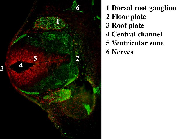
| Descripció | Antibody stain against Neurofilament (green) and Ki 67 (red) in a Mouse embryo at day 12.5 after fertilization. Shown is the dorsal root ganglion (green ellipsoid regions where cells express neurofilament) and the ventricular zone (red region where cells proliferate) as well as the neural tube with roof and floor plate. |
| Data | 04.05.2007 |
| Font | Treball propi |
| Autor | Hannes Röst |
Llicència
| Jo, el titular del copyright d'aquesta obra, l'allibero al domini públic. Això s'aplica a tot el món. En alguns països això pot no ser legalment possible, en tal cas: Jo faig concessió a tothom del dret d'usar aquesta obra per a qualsevol propòsit, sense cap condició llevat d'aquelles requerides per la llei. |
Historial del fitxer
Cliqueu una data/hora per veure el fitxer tal com era aleshores.
| Data/hora | Miniatura | Dimensions | Usuari/a | Comentari | |
|---|---|---|---|---|---|
| actual | 01:30, 2 nov 2007 | | 1.206 × 945 (607 Ko) | Hannes Röst | == Summary == {{Information |Description=Antibody stain against Neurofilament (green) and Ki 67 (red) in a Mouse embryo at day 12.5 after fertilization. Shown is the dorsal root ganglion (green ellipsoid regions where cells express neurofilament) and the |
| 00:03, 17 maig 2007 |  | 1.181 × 945 (149 Ko) | Hannes Röst | {{Information |Description=Antibody stain against Neurofilament (green) and Ki 67 (red) in a Mouse embryo. Shown is the neural tube development. |Source=self-made |Date=04.05.2007 |Author= User:Hannes Röst }} |
Ús del fitxer
La pàgina següent utilitza aquest fitxer:
Ús global del fitxer
Utilització d'aquest fitxer en altres wikis:
- Utilització a en.wikipedia.org
- Utilització a es.wikipedia.org
- Utilització a fr.wikipedia.org
- Utilització a gl.wikipedia.org
- Utilització a ja.wikipedia.org
- Utilització a pl.wikipedia.org