Fitxer:Unctional complex and pinocytotic vesicles - embryonic brain - TEM.jpg

Fitxer original (1.600 × 1.278 píxels, mida del fitxer: 861 Ko, tipus MIME: image/jpeg)
| Aquest fitxer i la informació mostrada a continuació provenen del dipòsit multimèdia lliure Wikimedia Commons. |
Resum
| Descripció |
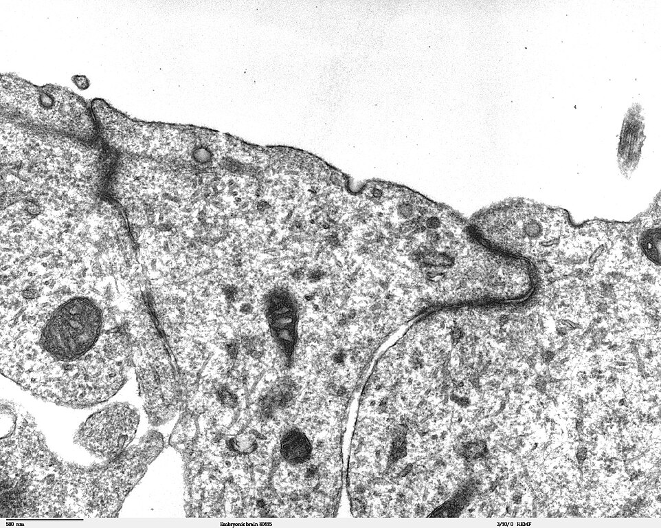
Transmission electron microscope image of a thin section cut through the developing brain tissue (telencephalic hemisphere) of an 11.5 day mouse embryo. This image of the luminal surface of the telencephalon, shows junctional complexes and pinocytotic vesicles. The junctional complex is divided into three types of junctions: 1) the most apical is the tight junction, which controls and/or restricts the movement of molecules across epithelial layers and helps maintain polarity, 2) the zonula adherens, which also includes the numerous actin filaments seen in the apical cytoplasm, and 3) the desmosome, which is a spot junction. The pinocytotic vesicles are formed from coated pits in the plasma membrane and are involved in endocytosis. JEOL 100CX TEM References: Marin-Padilla, M. (1985) "Early Vascularization of the Embryonic Cerebral Cortex: Golgi and Electron Microscope Studies", J. Comparative Neurology, 241:237-249 Marin-Padilla, M. and M. Amievo (1989) "Early Neurogenesis of the Mouse Olfactory Nerve: Golgi and Electron Microscope Studies", J. Comparative Neurology, 288:339-352 |
| Font | |
| Autor | Louisa Howard, Miguel Marin-Padilla |
| Permís (Com reutilitzar aquest fitxer) |
PD |
Llicència
| S'ha alliberat aquesta obra al domini públic pel seu autor Louisa Howard, Miguel Marin-Padilla. Això s'aplica a tot el món. En alguns països això pot no ser legalment possible, en tal cas: Louisa Howard, Miguel Marin-Padilla concedeix a tothom el dret d'usar aquesta obra per a qualsevol propòsit, sense cap condició llevat d'aquelles requerides per la llei.
|
Historial del fitxer
Cliqueu una data/hora per veure el fitxer tal com era aleshores.
| Data/hora | Miniatura | Dimensions | Usuari/a | Comentari | |
|---|---|---|---|---|---|
| actual | 23:06, 2 nov 2006 | | 1.600 × 1.278 (861 Ko) | Patho | {{Information |Description=Transmission electron microscope image of a thin section cut through the developing brain tissue (telencephalic hemisphere) of an 11.5 day mouse embryo. This higher magnification image of "Embryonic brain 80415", shows an area o |
Ús del fitxer
La pàgina següent utilitza aquest fitxer:
Ús global del fitxer
Utilització d'aquest fitxer en altres wikis:
- Utilització a bs.wikipedia.org
- Utilització a de.wikipedia.org
- Utilització a de.wikibooks.org
- Utilització a en.wikibooks.org
- Utilització a et.wikipedia.org
- Utilització a fr.wikipedia.org