Fitxer:Virus Replication.svg

Fitxer original (fitxer SVG, nominalment 462 × 426 píxels, mida del fitxer: 205 Ko)
| Aquest fitxer i la informació mostrada a continuació provenen del dipòsit multimèdia lliure Wikimedia Commons. |
Resum
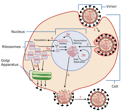
| Descripció | A diagram of influenza viral cell invasion and replication. |
| Data | |
| Font | Redrawn from w:Image:Virusreplication.png using Adobe Illustrator. |
| Autor | User:YK Times |
| Altres versions |
 |


|
Aquest fitxer SVG inclou text encapsulat que es pot traduir al vostre idioma utilitzant un editor que admeti SVG, un editor de text o l'eina de traducció de SVG. Per a més informació vegeu l'ajuda de traducció de fitxers SVG. |
Description from Scheme of Influenza A virus replication (NCBI): "A virion attaches to the host cell membrane via HA and enters the cytoplasm by receptor-mediated endocytosis (STEP 1), thereby forming an endosome. A cellular trypsin-like enzyme cleaves HA into products HA1 and HA2 (not shown). HA2 promotes fusion of the virus envelope and the endosome membranes. A minor virus envelope protein M2 acts as a ion channel thereby making the inside of the virion more acidic. As a result, the major envelope protein M1 dissociates from the nucleocapsid and vRNPs are translocated into the nucleus (STEP 2) via interaction between NP and cellular transport machinery. In the nucleus, the viral polymerase complexes transcribe (STEP 3a) and replicate (STEP 3b) the vRNAs. Newly synthesized mRNAs migrate to cytoplasm (STEP 4) where they are translated. Posttranslational processing of HA, NA, and M2 includes transportation via Golgi apparatus to the cell membrane (STEP 5b). NP, M1, NS1 (nonstructural regulatory protein - not shown) and NEP (nuclear export protein, a minor virion component - not shown) move to the nucleus (STEP 5a) where they bind freshly synthesized copies of vRNAs. The newly formed nucleocapsids migrate into the cytoplasm in a NEP-dependent process and eventually interact via M1 with a region of the cell membrane where HA, NA and M2 have been inserted (STEP 6). Then the newly synthesized virions bud from infected cell (STEP 7). NA destroys the sialic acid moiety of cellular receptors, thereby releasing the progeny virions."
Llicència

|
S'autoritza la còpia, la distribució i la modificació d'aquest document sota els termes de la llicència de documentació lliure GNU versió 1.2 o qualsevol altra versió posterior que publiqui la Free Software Foundation; sense seccions invariants, ni textos de portada, ni textos de contraportada. S'inclou una còpia d'aquesta llicència en la secció titulada GNU Free Documentation License. |
| Aquest fitxer està subjecte a la llicència de Creative Commons Reconeixement i Compartir Igual 3.0 No adaptada. | ||
| ||
| Aquest avís de llicència s'ha afegit a aquest fitxer d'acord amb l'actualització de la llicència GFDL. |
- Sou lliure de:
- compartir – copiar, distribuir i comunicar públicament l'obra
- adaptar – fer-ne obres derivades
- Amb les condicions següents:
- reconeixement – Heu de donar la informació adequada sobre l'autor, proporcionar un enllaç a la llicència i indicar si s'han realitzat canvis. Podeu fer-ho amb qualsevol mitjà raonable, però de cap manera no suggereixi que l'autor us dóna suport o aprova l'ús que en feu.
- compartir igual – Si modifiqueu, transformeu, o generareu amb el material, haureu de distribuir les vostres contribucions sota una llicència similar o una de compatible com l'original
| Anotacions | Aquesta imatge té anotacions: Vegeu-les a Commons |
Historial del fitxer
Cliqueu una data/hora per veure el fitxer tal com era aleshores.
| Data/hora | Miniatura | Dimensions | Usuari/a | Comentari | |
|---|---|---|---|---|---|
| actual | 04:54, 6 març 2007 | | 462 × 426 (205 Ko) | YK Times | {{Information |Description=A diagram of influenza viral cell invasion and replication. |Source=Redrawn from w:Image:Virusreplication.png using Adobe Illustrator. |Date=March 5, 2007 |Author= User:YK Times |Permission= |other_versions=[[:w:Image:V |
Ús del fitxer
La pàgina següent utilitza aquest fitxer:
Ús global del fitxer
Utilització d'aquest fitxer en altres wikis:
- Utilització a bg.wikipedia.org
- Utilització a bn.wikipedia.org
- Utilització a br.wikipedia.org
- Utilització a cs.wikipedia.org
- Utilització a da.wikipedia.org
- Utilització a de.wikipedia.org
- Utilització a el.wikipedia.org
- Utilització a en.wikipedia.org
- Orthomyxoviridae
- Amantadine
- Viral replication
- Viral life cycle
- Viral entry
- File:Virusreplication.png
- User:YK Times/Graphic Lab/examples
- Wikipedia:Graphics Lab/Images to improve/Archive/Mar 2007
- Viral shedding
- Template:Influenza virus life cycle
- Viral neuraminidase
- Tilapia tilapinevirus
- User:Zoe.gum/sandbox
- Wikipedia talk:WikiProject Viruses/Archive 4
- User:Anicm1/sandbox
- Utilització a en.wikibooks.org
- Utilització a es.wikipedia.org
- Utilització a fa.wikipedia.org
- Utilització a fr.wikipedia.org
- Utilització a hy.wikipedia.org
- Utilització a id.wikipedia.org
- Utilització a it.wikipedia.org
- Utilització a ja.wikipedia.org
- Utilització a kk.wikipedia.org
- Utilització a ko.wikipedia.org
Vegeu més usos globals d'aquest fitxer.